


IMPORTANT NOTICE: The information presented in this article is for educational and informational purposes only and does not constitute medical advice. Cancer is a complex disease involving multiple interconnected mechanisms, including genetic mutations, metabolic dysfunction, immune dysregulation, environmental exposures, and lifestyle factors. The sodium-potassium-inflammation link discussed herein represents one aspect of a multifaceted disease process. This content is not intended to diagnose, treat, cure, or prevent any disease. Always consult with qualified healthcare professionals—including oncologists, nutritionists, and integrative medicine practitioners—before making any changes to diet, supplements, or treatment protocols. Cancer management should always be personalized and supervised by experienced medical professionals.
Introduction: Rethinking Cancer’s Origins
Cancer remains one of the most feared diagnoses of our time. When patients receive this diagnosis and ask their oncologists, “Why me?” the response often points to genetics—”It’s in your genes,” “You had bad luck,” or “It’s a random mutation.” Yet epidemiological data tell a different story.
The numbers are staggering: current statistics indicate that 1 in 2 men and 1 in 3 women will receive a cancer diagnosis in their lifetime (American Cancer Society, 2023). This represents a dramatic increase from previous generations—a trend that cannot be explained by genetics alone, as our gene pool has not changed meaningfully over the past century.
The prevailing paradigm in orthodox oncology has long treated cancer as primarily a genetic disease—a “mutational” condition where DNA damage triggers uncontrolled cell division. Billions of dollars in research have been directed toward understanding and targeting these mutations. However, a growing body of evidence suggests this model may be incomplete.
The Metabolic Revolution
We now understand that cancer is fundamentally a metabolic disease (Seyfried & Shelton, 2010). The work of Nobel laureate Otto Warburg nearly a century ago demonstrated that cancer cells undergo a metabolic shift—switching from oxidative phosphorylation to aerobic glycolysis (the “Warburg effect”), consuming glucose at dramatically increased rates. This metabolic reprogramming appears to be a driver, not merely a consequence, of malignant transformation.
The evidence for this is compelling. In landmark experiments, researchers removed the nuclei from cancerous cells and inserted them into normal cells. The result was that nothing happened—the normal cytoplasm remained normal. However, when healthy nuclei were inserted into the cytoplasm of cancerous cells, those cells became cancerous (McKinnell et al., 1969). This demonstrated that the “cancer program” resides not in the nucleus but in the cytoplasm—in the metabolic machinery of the cell. This finding has profound implications for how we understand and approach cancer treatment.
Beyond the Nuclear Dogma
Despite such findings, why hasn’t metabolic oncology been adopted by mainstream medicine? Follow the money:
The average cancer patient generates approximately $150,000–$300,000 in treatment costs over their disease course (Mariotto et al., 2011)
The global oncology drug market exceeds $200 billion annually (IQVIA Institute, 2023)
Conventional treatments—chemotherapy, radiation, surgery, immunotherapy—represent a multi-trillion-dollar industry
In contrast, telling patients to modify their diet, exercise, sleep, and lifestyle generates no patentable drugs and no multi-billion-dollar revenues. The economic incentives for maintaining the status quo are enormous and cannot be ignored.
The Metabolic Context of Cancer
Cancer cells differ from healthy cells in multiple fundamental ways:
Metabolic reprogramming: Cancer cells preferentially use fermentation (glycolysis) rather than oxidative phosphorylation for energy production, even in the presence of oxygen (Warburg, 1956)
Oxidative stress: Environmental toxins—including industrial chemicals, microplastics, household products, herbicides, pesticides, and electromagnetic fields (EMF)—generate reactive oxygen species (ROS) that damage cellular components
Inflammation: Chronic inflammation is now recognized as a hallmark of cancer development (Hanahan & Weinberg, 2011)
Immune dysregulation: Cancer cells develop mechanisms to evade immune surveillance
Growth factor independence: Cancer cells acquire the ability to proliferate without external growth signals
The Inflammation-Cancer Connection
The relationship between inflammation and cancer has been extensively documented. The work of Professor Mahin Khatami has been particularly instrumental in understanding this association (Khatami, 2009, 2014). Chronic inflammation creates an environment of tissue damage, oxidative stress, and cellular proliferation that promotes malignant transformation.
The Historical Foundation: Schweitzer and Gerson
The story of the sodium-potassium ratio in cancer begins with a remarkable historical encounter.
Dr. Albert Schweitzer (1875–1965), the renowned physician, philosopher, and organist, founded a hospital in Lambaréné, Gabon (then French Equatorial Africa) during World War I. In his decades of service there, he made a striking observation: cancer was virtually unknown among the indigenous population who maintained traditional diets and lifestyles.
However, as the European diet was introduced—characterized by refined foods, preserved meats, and increased salt consumption—cancer began to appear among local people. Schweitzer reflected deeply on what had changed in their environment and nutrition.
Dr. Max Gerson (1881–1959), a German-born physician, had developed a dietary approach to treating cancer that proved so controversial he was forced to flee Nazi Germany. Gerson’s protocol emphasized:
Organic, plant-based foods
Fresh vegetable juices
High potassium intake
Low sodium intake
Liver-supporting nutrients
In a conversation recorded in Schweitzer’s biography (Schweitzer & Joy, 1966), Schweitzer and Gerson discussed the cancer phenomenon. Schweitzer shared his observation that cancer was absent among traditional Africans. After reflection, he hypothesized that excessive salt might be the culprit.
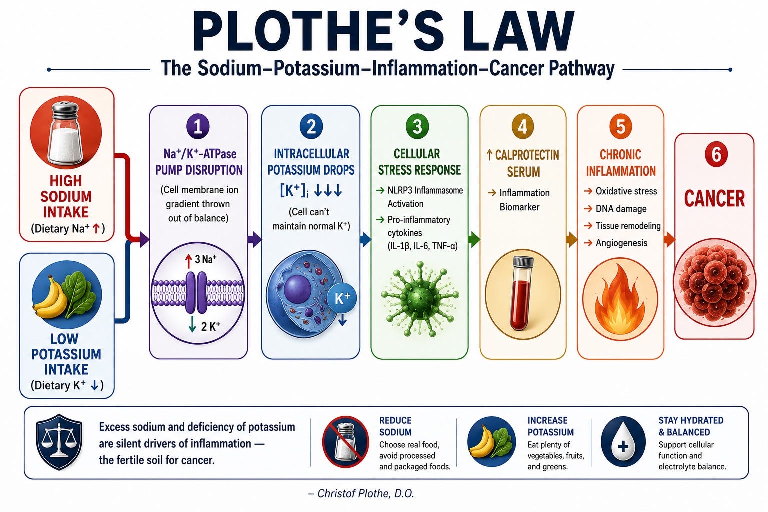
Gerson agreed but refined the analysis: it wasn’t just the presence of salt but the imbalance—high sodium combined with insufficient potassium created the cancer-permissive environment. Their rationale was that once too much sodium enters cells, it displaces potassium, creating intracellular inflammation measurable by elevated calprotectin levels (a marker of neutrophil activity and intestinal inflammation).
Basic Cellular Biology: The Sodium-Potassium Pump
To understand the significance of the sodium-potassium ratio, we must first understand basic cellular physiology.
The Electrochemical Gradient
In a healthy cell:
Extracellular space: High sodium (Na⁺), low potassium (K⁺)
Intracellular space: Low sodium (Na⁺), high potassium (K⁺)
This gradient is maintained by the Na⁺/K⁺-ATPase pump, a membrane protein that:
Pumps 3 sodium ions out of the cell
Pumps 2 potassium ions into the cell
Consumes 1 ATP per cycle
This pump consumes approximately 30–50% of all cellular ATP production, making it one of the most energy-demanding processes in the body. Its proper function is essential for:
Maintaining membrane potential
Regulating cell volume
Driving secondary active transport
Enabling nerve impulse transmission
Supporting muscle contraction
When the Gradient Fails
Several factors can disrupt the sodium-potassium gradient in cancer cells:
Dysregulated ion pumps: The Na⁺/K⁺-ATPase may be downregulated or dysfunctional in cancer cells
Energy deficits: The Warburg effect (aerobic glycolysis) reduces ATP availability
Hypoxia: Low oxygen levels impair ATP production
Altered membrane channels: Cancer cells may overexpress sodium channels (ENaC) while suppressing potassium channels
The consequences of this disruption are profound:
Apoptosis inhibition: Low intracellular potassium inhibits caspase activation, preventing programmed cell death (Bortner & Cidlowski, 1998)
Proliferation promotion: High intracellular sodium activates growth-promoting pathways (MAPK/ERK)
Migration enhancement: Altered ion balance aids cell migration and metastasis
pH regulation: Sodium-hydrogen exchangers help cancer cells maintain pH despite lactic acid production
The Evidence: Jansson’s Seminal Review
The foundational work connecting potassium, sodium, and cancer comes from B. Jansson’s comprehensive review (Jansson, 1996).
Key Findings
Jansson (1996) found that:
Carcinogenic agents decrease cellular potassium and increase cellular sodium
Anticarcinogenic agents have the opposite effect—increasing potassium and decreasing sodium
This relationship was consistently observed across all agents studied where data were available
Specific Agents and Their Effects
Dietary Carcinogenic Agents (increase Na⁺, decrease K⁺):
Sodium (excess salt)
Cadmium
Fat
Cholesterol
Calories (excess)
Alcohol
Dietary Anticarcinogenic Agents (increase K⁺, decrease Na⁺):
Potassium
Vitamins A, C, and D
Selenium
Fiber
Other Carcinogenic and Anticarcinogenic Agents
Jansson (1996) further demonstrated that the pattern extends beyond diet:
Dimethylhydrazine (a cancer-causing drug) increases sodium and decreases potassium in cells
Indomethacin (an anticancer agent in some contexts) has the opposite effect
Aging and Cancer
With aging:
Potassium leaves cells
Sodium enters cells
Cancer rates increase
This age-related ionic shift may contribute to the increased cancer risk observed in older populations.
Disease Associations
Jansson (1996) also noted:
Patients with hyperkalemic diseases (e.g., Parkinson’s disease, Addison’s disease) have reduced cancer rates
Patients with hypokalemic diseases (e.g., alcoholism, obesity, stress states) have increased cancer rates
The Ratio Matters Most
Perhaps most importantly, Jansson (1996) concluded that the ratio between intracellular potassium and sodium was of greater importance than either element individually. The relationship between these concentrations and cancer rates is hyperbolic rather than linear—meaning that two independent carcinogenic agents can be synergistic rather than additive in their effects.
The Evolutionary Perspective
Jansson (1990) placed the sodium-potassium imbalance in evolutionary context:
The Paleolithic Baseline
During human evolution:
Potassium intake: Approximately 10g/day from fruits, vegetables, roots, and nuts
Sodium intake: Approximately 1-2g from natural sources
The Modern Inversion
Today, the situation is reversed:
Sodium intake: >10g/day (mostly from processed foods and added salt)
Potassium intake: Only 1-2g/day
The dietary K/Na ratio has been reduced by a factor of about 20 from Paleolithic times to modern times. When comparing modern people in industrialized areas with the Yanomamo Indians of South America—who eat no added salt but consume potassium-rich cooking bananas—the factor may be 100 to 200 (Jansson, 1990).
The Unresolved Adaptation
Humans evolved to:
Conserve sodium from a sodium-poor environment
Excrete potassium from a potassium-rich environment
We have not yet evolutionarily adapted to today’s high-sodium, low-potassium diet. This mismatch has created increased rates of multiple diseases, including cancer.
The Recommended Ratio
Jansson (1990) recommended:
Dietary K/Na ratio should be well above 1
Preferably 5 or higher
Cellular K/Na ratio should be above 10
The Mechanism: Sodium, Potassium, and Inflammation
The Inflammasome Connection
The critical mechanistic link between sodium-potassium imbalance and cancer involves the NLRP3 inflammasome.
Muñoz-Planillo et al. (2013) demonstrated that potassium efflux is the common trigger for NLRP3 inflammasome activation. When intracellular potassium drops, NLRP3 oligomerizes and activates, leading to cleavage of pro-IL-1β into active IL-1β—one of the most potent pro-inflammatory cytokines in the body.
Binger et al. (2015) showed that high salt conditions activate macrophages via the transcription factor NFAT5 (TonEBP), driving them toward a pro-inflammatory state.
Zhang et al. (2015) identified a distinct macrophage phenotype—M(Na)—induced by high salt conditions, characterized by:
Increased IL-1β production
Elevated TNF-α
Enhanced IL-6
NLRP3/caspase-1 pathway activation
The Th17 Autoimmune Axis
Kleinewietfeld et al. (2013) demonstrated that high salt conditions:
Induce naïve CD4⁺ T cells to differentiate into pathogenic Th17 cells
Do this via SGK1 (serum/glucocorticoid-regulated kinase 1)
SGK1 is induced by osmotic stress from high sodium
Th17 cells are implicated in multiple sclerosis, rheumatoid arthritis, and inflammatory bowel disease
The S100A8/A9-Calprotectin Pathway
This is where the connection to cancer becomes concrete.
Calprotectin (S100A8/A9) is a heterodimer of two calcium-binding proteins released from:
Neutrophils
Monocytes
Macrophages
Epithelial cells during inflammation
Vogl et al. (2007) demonstrated that S100A8 and S100A9 are endogenous activators of Toll-like receptor 4 (TLR4), promoting lethal endotoxin-induced shock. TLR4 activation triggers NF-κB, which drives inflammatory cytokine production.
Ghavami et al. (2008) showed that S100A8/A9 promotes tumor cell growth via:
RAGE (Receptor for Advanced Glycation End-products) ligation
MAP kinase pathway activation
The STAT3 Connection
Turovskaya et al. (2008) found that in colitis-associated cancer:
S100A8/A9 activates both TLR4 and RAGE
This converges on STAT3 phosphorylation
Constitutively active STAT3 is found in over 70% of human cancers
STAT3 drives:
Cyclin D1 and c-Myc (cell cycle progression)
Bcl-2 and Bcl-xL (anti-apoptotic proteins)
VEGF (angiogenesis)
MMP-2 and MMP-9 (invasion and metastasis)
Myeloid-Derived Suppressor Cells (MDSCs)
Sinha et al. (2008) revealed that S100A8/A9:
Acts as a chemoattractant for MDSCs
Promotes their expansion in bone marrow
Facilitates their migration to peripheral tissues
MDSCs suppress CD8⁺ T cells via arginase-1, iNOS, and ROS production
This creates an immunosuppressive microenvironment for tumors
The Complete Chain
High Na⁺ / Low K⁺ Ratio
→ Membrane depolarization + NLRP3 derepression
→ S100A8/A9 release into serum (serum calprotectin)
→ TLR4 + RAGE activation → NF-κB + STAT3
→ Genomic Instability (AID/APOBEC mutations)
→ MDSC Recruitment (immune suppression)
→ COX-2/PGE₂ Feedback Loop (sustained inflammation)
→ Proliferative Signaling (Cyclin D1, c-Myc)
→ Anti-apoptotic State
→ CARCINOGENESIS
The Three Pillars of Investigation: A Monitoring Framework
Based on the scientific evidence, a comprehensive monitoring approach for cancer patients and those at risk should include three key biomarkers assessed every 6 weeks:
Pillar 1: 24-Hour Urinary Sodium Test
This test provides an objective measure of daily sodium consumption, free from the recall bias inherent in dietary questionnaires.
The Evidence:
A landmark prospective cohort study by Wang et al. (2024), published in Clinical and Translational Gastroenterology, analyzed 434,372 individuals from the UK Biobank over a median follow-up of 13.8 years. The findings were striking:
Each 1-gram increase in estimated 24-hour urinary sodium excretion corresponded to a 12% increased risk of pancreatic cancer (HR = 1.12, 95% CI: 1.03, 1.22)
Participants in the highest quartile of sodium excretion had a 38% higher risk (HR = 1.38, 95% CI: 1.21, 1.58) compared to the lowest quartile
1,765 cases of pancreatic cancer were detected during the study period (Wang et al., 2024)
The study authors noted: “These results propose an association between elevated sodium consumption and a heightened risk of pancreatic cancer” (Wang et al., 2024). This builds on prior research linking high sodium intake to gastric cancer (D’Elia et al., 2012) and renal cell carcinoma (Deckers et al., 2014).
The Salt Substitute Evidence:
The Salt Substitute and Stroke Study (SSaSS), a landmark cluster-randomized trial involving 20,995 participants over 4.7 years, evaluated the effects of replacing regular salt (100% sodium chloride) with a potassium-enriched salt substitute (75% sodium chloride, 25% potassium chloride) (Neal et al., 2021; SSaSS Investigators, 2025).
While the primary analysis focused on cardiovascular outcomes, a post-hoc cancer analysis revealed:
Trend toward protection against fatal stomach cancer: Rate Ratio 0.66 (95% CI: 0.44 to 1.00)
Composite outcome (stomach, colorectal, liver, and lung cancers): Rate Ratio 0.76 (95% CI: 0.62 to 0.92)
Mean 24-hour urinary sodium excretion measured in 614 participants was 4.3 g/day, equating to daily salt consumption of about 11.9 g/day—far exceeding WHO recommendations of <5 g/day (SSaSS Investigators, 2025)
The study authors concluded: “Trends toward protection against fatal and non-fatal stomach cancer align with the observational epidemiology and warrant further investigation” (SSaSS Investigators, 2025).
Practical Guidance:
24-hour urine collection is the gold standard for assessing sodium intake
Target: <100 mmol (2.3 g) sodium per 24 hours (WHO recommendation)
Results can guide dietary sodium reduction strategies
Pillar 2: Intracellular Potassium Measurement
Assessing intracellular potassium status is more challenging than measuring serum potassium, as serum levels do not accurately reflect intracellular stores.
The Evidence:
A recent breakthrough study by Yang et al. (2025), published in the Journal of the American Chemical Society, developed the first highly selective DNAzyme-based sensor for intracellular potassium imaging. Key findings include:
Progressive decline in intracellular K⁺ levels in breast cancer cells with more advanced progression states
Elevated extracellular K⁺ interferes with anticancer compound efficacy (ML133 and Amiodarone), suggesting a role in chemoresistance
Kir2.1 channel blockade restored treatment sensitivity, presenting a potential strategy to overcome chemoresistance in aggressive cancers
The authors stated: “These findings underscore the role of K⁺ homeostasis in tumor progression and support further exploration of ion-channel-targeted cancer therapies” (Yang et al., 2025).
This builds on earlier work by Wang et al. (2021), who developed Ratiometric Potassium Sensor 1 (RPS-1) and found elevations in basal intracellular K⁺ in metastatic breast cancer cell lines versus normal breast cells, suggesting that potassium dynamics are complex and context-dependent.
Huang et al. (2025) developed a cell surface biosensor for single-cell ion sequencing (Ion-seq) to monitor potassium homeostasis in clinical colorectal cancer samples. The study noted that increased extracellular K⁺ during tumor development disrupts infiltrating T cell differentiation, compromising T cell-based tumor immunotherapy efficacy (Huang et al., 2025).
Practical Guidance:
Standard serum potassium tests do not accurately reflect intracellular levels
Emerging technologies such as DNAzyme-based sensors and single-cell ion sequencing are advancing our ability to assess intracellular potassium
Red blood cell potassium measurements may provide a more accurate reflection of intracellular status
Focus on dietary strategies to increase potassium intake through whole foods
Pillar 3: Calprotectin (Serum + Fecal)
Calprotectin (S100A8/A9) is a marker of inflammation that is more specific to the innate immune/inflammasome axis than CRP.
The Evidence:
Vogl et al. (2007) established that S100A8 and S100A9 are endogenous activators of Toll-like receptor 4 (TLR4), promoting lethal inflammation. This positions calprotectin as a key mediator at the intersection of inflammation and carcinogenesis.
Turovskaya et al. (2008) demonstrated that S100A8/A9 plays essential roles in colitis-associated carcinogenesis through RAGE and TLR4 activation, converging on STAT3 phosphorylation.
Ichikawa et al. (2011) showed that S100A8/A9 activates key genes and pathways in colon tumor progression and is necessary for tumor progression in multiple models—blocking it stopped tumor growth.
Sinha et al. (2008) revealed that S100A8/A9 promotes the accumulation of myeloid-derived suppressor cells (MDSCs), which suppress CD8⁺ T cell function, contributing to immune evasion.
Fecal calprotectin is already used clinically as a screening adjunct for colorectal inflammation and cancer. Serum calprotectin tracks the systemic inflammatory component.
Practical Guidance:
Measure both fecal and serum calprotectin every 6 weeks
Fecal calprotectin reflects intestinal inflammation
Serum calprotectin reflects systemic inflammatory burden
Elevations may indicate active inflammatory processes that could promote cancer progression
Normalization of calprotectin can serve as a marker of successful anti-inflammatory intervention
Monitoring Schedule Summary
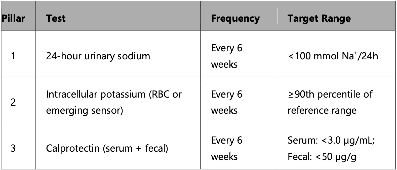
Rationale for 6-week intervals:
Allows sufficient time for dietary and lifestyle interventions to produce measurable changes
Balances clinical utility with patient burden
Matches typical clinical monitoring schedules for cancer patients
Enables tracking of trends and early identification of concerning changes
Investigated Cancers Linked to This Pathway
Colorectal Cancer
The best-established link. Fecal calprotectin is already used clinically as a screening adjunct, and serum calprotectin tracks the systemic inflammatory component.
Ichikawa et al. (2011) showed S100A8/A9 was necessary for colon tumor progression in multiple models. Blocking it stopped tumor growth.
Gastric Cancer
Given the H. pylori → salt → inflammation connection, gastric cancer is the natural endpoint. S100A8/A9 is upregulated in gastric cancer tissue, and serum levels correlate with tumor stage (Yong & Moon, 2007).
Pancreatic Cancer
Wang et al. (2024) provided compelling prospective evidence linking sodium intake to pancreatic cancer risk. Pancreatic cancer is notoriously immunosuppressed, with dense MDSC infiltrates. Serum S100A8/A9 is a major MDSC chemoattractant in pancreatic cancer (Basso et al., 2014).
Breast and Lung Cancer
The S100A8/A9 pre-metastatic niche concept applies here. Before tumor cells metastasize, S100A8/A9 from the primary tumor (or systemic inflammation) primes target organs by recruiting MDSCs and creating an immunosuppressive microenvironment (Hiratsuka et al., 2006).
Yang et al. (2025) demonstrated progressive decline in intracellular K⁺ levels in breast cancer cells with more advanced progression states, and Wang et al. (2021) found elevated basal intracellular K⁺ in metastatic breast cancer cell lines—suggesting complex, context-dependent potassium dynamics.
Dietary Strategies to Correct the Imbalance
Increase Potassium-Rich Foods
Potassium helps maintain normal cell membrane potential and supports apoptosis.
Top Sources:
Leafy greens (spinach, Swiss chard)
Avocados (highest K⁺ content among fruits)
Bananas, oranges, cantaloupe
Potatoes (with skin), sweet potatoes
Beans (white beans, lentils)
Coconut water (natural electrolyte)
Beets and beet greens
Beet root deserves special mention as an exceptional potassium source with additional benefits for nitric oxide production and vascular health.
Reduce Excess Sodium
High sodium promotes proliferation and metastasis.
Recommendations:
Avoid processed meats, canned soups, chips, and fast food
Use Himalayan pink salt or sea salt in moderation (contains trace minerals)
Flavor foods with herbs and spices instead of salt
Read food labels carefully
Consider potassium-enriched salt substitutes (75% sodium chloride, 25% potassium chloride)
The Salt Substitute Evidence
The SSaSS trial (Neal et al., 2021; SSaSS Investigators, 2025) demonstrated that switching to potassium-enriched salt substitutes:
Reduced stroke rate by 14% (RR = 0.86)
Reduced major cardiovascular events by 13% (RR = 0.87)
Reduced all-cause mortality by 12% (RR = 0.88)
Showed trends toward cancer protection, particularly for stomach cancer
Magnesium Synergy
Magnesium supports Na⁺/K⁺-ATPase pump function. Low magnesium worsens potassium deficiency.
Sources:
Pumpkin seeds, almonds, cashews
Dark chocolate (85%+ cocoa)
Spinach, black beans
Alkaline Diet Considerations
Cancer cells thrive in acidic environments; potassium-rich foods are often alkaline-forming.
Foods to Favor:
Citrus fruits (lemons, limes—alkalizing despite acidity)
Cruciferous vegetables (broccoli, kale)
Apple cider vinegar (balances pH)
Herbal and Natural Support
Herbs That Modulate Na⁺/K⁺ Pumps
Hawthorn (Crataegus): Supports Na⁺/K⁺-ATPase function (Parikh et al., 2015)
Ginseng (Panax ginseng): May enhance ion pump activity (Chen et al., 2009; Hu et al., 2016) .
Astragalus: Regulates cellular metabolism (Huang et al., 2012; Huang et al.) 2017) .
Potassium-Sparing Diuretics (Natural)
Dandelion root: Mild diuretic without depleting potassium ((EMA, 2008; EMA, 2020; Hameed,2023) .
Nettle leaf: Rich in potassium, supports kidney function (Salman, 2026)
Herbs That Lower Intracellular Sodium
Celery seed: May help excrete excess sodium (Escobedo-Gutiérrez, 2026; Liu, 2025)
Hibiscus tea: Mild diuretic and sodium-lowering effects (Konaté, 2024)
Adaptogens for Cellular Stress
Ashwagandha: May regulate ion channels and reduce oxidative stress (Fanibunda, 2025; Singh,2026).
Reishi mushroom: Supports immune function and cellular homeostasis (Chen, 2023; Zhang, 2025).
Clinical Implications
Calprotectin as a Biomarker
Serum calprotectin may be more valuable than CRP for monitoring cancer risk because:
CRP is produced by the liver in response to IL-6
Calprotectin is released at the actual site of immune activation
Calprotectin is more specific to the innate immune/inflammasome axis that drives carcinogenesis
In cancer patients, measuring calprotectin every 6 weeks can help track disease progression and inflammatory status.
The Feedback Loop Challenge
Once the COX-2/PGE₂/S100A8/A9 feedback loop is running, it can become self-sustaining. This means:
Fixing electrolytes alone may not be sufficient
Targeted interventions may be needed to break the cycle
These could include:
Low-dose aspirin
COX-2 selective inhibitors (under medical supervision)
S100A8/A9-blocking biologics (in development)
Practical Recommendations
Test don’t guess: A 24-hour urine sodium test provides objective data on salt excretion
Replace your salt: Use potassium chloride salt substitutes
Eat the rainbow: Prioritize potassium-rich whole foods
Minimize processed foods: This is where most dietary sodium comes from
Consider magnesium: It’s essential for pump function
Monitor regularly: Every 6 weeks for the three pillars
Work with professionals: Cancer is complex; any dietary changes should be made in consultation with healthcare providers
Critical Considerations and Cautions
The Complexity of Cancer
Cancer is multifactorial. The sodium-potassium-inflammation link is one important mechanism among many:
Genetic mutations
Hormonal factors
Immune function
Environmental exposures
Other dietary factors
Physical activity
Sleep quality
Stress management
Potassium Supplementation
Do NOT take high-dose potassium supplements without medical supervision—but consider it when investigations prove that this can be essential for you as it can cause:
Dangerous arrhythmias
Cardiac arrest
Muscle paralysis
The Role of Sodium
Sodium is essential for life. The goal is not elimination but balance:
Prioritize whole foods
Avoid processed foods
Use salt substitutes
Monitor intake
Individual Variation
People vary widely in:
Salt sensitivity
Baseline potassium status
Kidney function
Overall health status
The Need for Professional Guidance
Always work with:
An experienced holistic oncologist
A registered dietitian
An integrative medicine practitioner
Conclusion
The sodium-potassium ratio represents one of the most underestimated factors in cancer development and progression. The evidence is compelling:
Evolutionary mismatch: Our modern diet has inverted the sodium-potassium ratio from our evolutionary baseline (Jansson, 1990)
Cellular consequences: High sodium/low potassium disrupts the Na⁺/K⁺ pump, creates membrane depolarization, and drives inflammation (Jansson, 1996)
Inflammatory cascade: Low intracellular potassium triggers NLRP3 inflammasome activation, S100A8/A9 release, TLR4/RAGE activation, and NF-κB/STAT3 signaling (Muñoz-Planillo et al., 2013; Vogl et al., 2007; Turovskaya et al., 2008)
Cancer promotion: This inflammatory state creates genomic instability, immunosuppression, and a proliferative environment (Sinha et al., 2008; Ichikawa et al., 2011)
Dietary intervention: Restoring the potassium-sodium balance through diet—with potassium-rich whole foods and reduced processed salt—may be one of the most fundamental anti-cancer interventions (SSaSS Investigators, 2025)
Monitoring: Regular assessment of 24-hour urinary sodium, intracellular potassium, and calprotectin every 6 weeks provides objective data to guide interventions (Wang et al., 2024; Yang et al., 2025; Huang et al., 2025)
The historical insights of Schweitzer and Gerson, confirmed by modern research, point to a simple but profound truth: the balance between potassium and sodium in our bodies may be a key determinant of whether inflammation fuels cancer or health prevails.
References
1. Basso, D., Fogar, P., Plebani, M., & Padoan, A. (2014). S100A8/A9 in pancreatic cancer. Clinica Chimica Acta, 436, 85-90.
2. Binger, K. J., Gebhardt, M., Heinig, M., Rintisch, C., Schroeder, A., Neuhofer, W., ... & Titze, J. (2015). High salt reduces the activation of IL-4- and IL-13-stimulated macrophages. Journal of Clinical Investigation, 125(11), 4223-4238.
3. Bortner, C. D., & Cidlowski, J. A. (1998). A necessary role for cell shrinkage in apoptosis. Biochemical Pharmacology, 56(12), 1549-1559.
4. Chen, R. J., Chung, T. Y., Li, F. Y., Lin, N. H., & Tzen, J. T. C. (2009). Effect of sugar positions in ginsenosides and their inhibitory potency on Na⁺/K⁺-ATPase activity. Acta Pharmacologica Sinica, 30(1), 61–69. https://doi.org/10.1038/aps.2008.6
5. Chen, S. N., et al. (2023). Efficacy and safety of Reishi β-glucan in healthy adults: A randomized, double-blinded, placebo-controlled clinical trial. Foods, 12(4), 659–672. https://doi.org/10.3390/foods12040659
6. D’Elia, L., Rossi, G., Ippolito, R., Cappuccio, F. P., & Strazzullo, P. (2012). Habitual salt intake and risk of gastric cancer: A meta-analysis of prospective studies. Clinical Nutrition, 31(4), 489-498.
7. Deckers, I. A. G., van den Brandt, P. A., van Engeland, M., Soetekouw, P. M. M. B., Baldewijns, M. M. L. L., Goldbohm, R. A., & Schouten, L. J. (2014). Long-term dietary sodium, potassium and fluid intake; exploring potential novel risk factors for renal cell cancer in the Netherlands Cohort Study on diet and cancer. British Journal of Cancer, 110(3), 797-801.
8. Escobedo-Gutiérrez, M. d. J., Cortez-Navarrete, M., Martínez-Abundis, E., & Pérez-Rubio, K. G. (2026). Effect of Celery Seed (Apium graveolens L.) Administration on the Components of Metabolic Syndrome, Insulin Sensitivity, and Insulin Secretion: A Clinical Trial. Pharmaceuticals, 19(1), 110. https://doi.org/10.3390/ph19010110
9. European Medicines Agency (EMA). (2008). Final assessment report on Taraxacum officinale Weber ex Wigg., folium (EMA/HMPC/579634/2008). https://www.ema.europa.eu/en/documents/herbal-report/final-assessment-report-taraxacum-officinale-weber-ex-wigg-folium_en.pdf
10. European Medicines Agency (EMA). (2020). Assessment report on Taraxacum officinale F.H. Wigg., radix (EMA/HMPC/475725/2020). https://www.ema.europa.eu/en/documents/herbal-report/draft-assessment-report-taraxacum-officinale-fh-wigg-radix-first-version_en.pdf
11. Fanibunda, S. E., et al. (2025). Withania somnifera regulates mitochondrial biogenesis and energetics in rat cortical neurons: Role of BDNF and SIRT1. Journal of Neuroinflammation, 22, 04920–04927. https://doi.org/10.1007/s12035-025-04920-7
12. Ghavami, S., Rashedi, I., Dattilo, B. M., Eshraghi, M., Chazin, W. J., Hashemi, M., ... & Los, M. (2008). S100A8/A9 at low concentration promotes tumor cell growth via RAGE ligation and MAP kinase-dependent pathway. Journal of Leukocyte Biology, 83(6), 1484-1492.
13. Hanahan, D., & Weinberg, R. A. (2011). Hallmarks of cancer: The next generation. Cell, 144(5), 646-674.
14. Hiratsuka, S., Watanabe, A., Aburatani, H., & Maru, Y. (2006). Tumour-mediated upregulation of chemoattractants and recruitment of myeloid cells predetermines lung metastasis. Nature Cell Biology, 8(12), 1369-1375.
15. Hu, G., Zong, R. Y., & Shao, C. J. (2016). Effects of ginsenosides from stems and leaves on activities of Na⁺, K⁺-ATPase, Ca²⁺-ATPase and Mg²⁺-ATPase in rabbit cerebrum in vitro. Acta Pharmacologica Sinica, 9(6). http://www.chinaphar.com/article/view/8680
16. Huang Z, Shi X, Yang Y, Ma W, Li H, Jin Z, Feng Y, Luo C, Zheng L, Du Z, Liu C, Liu C, Liu Y, Wu Q, Liu L, Peng R, Tan W. Cell Surface Ion-Seq: Potassium Ion Monitoring in the Colorectal Cancer Cellular Microenvironment Based on Split G‑quadruplex Probe. ACS Cent Sci. 2025 Dec 5;11(12):2375-2384. doi: 10.1021/acscentsci.5c01472. PMID: 41473793; PMCID: PMC12746146.
17. Huang, X. P., Ding, H., Lu, J. D., Tang, Y. H., Deng, B. X., & Deng, C. Q. (2012). Astragalus extract alleviates nerve injury after cerebral ischemia by improving energy metabolism and inhibiting apoptosis. Biological & Pharmaceutical Bulletin, 35(4), 449–454. https://pubmed.ncbi.nlm.nih.gov/22466546/
18. Huang, X. P., Qiu, Y. Y., Wang, B., Ding, H., Tang, Y. H., Zeng, R., & Deng, C. Q. (2017). Combination of total Astragalus extract and total Panax notoginseng saponins strengthened the protective effects on brain damage through improving energy metabolism and inhibiting apoptosis after cerebral ischemia-reperfusion in mice. Chinese Journal of Integrative Medicine, 23(6), 445–452. https://doi.org/10.1007/s11655-015-1965-0
19. Ichikawa, M., Williams, R., Wang, L., Vogl, T., & Srikrishna, G. (2011). S100A8/A9 activate key genes and pathways in colon tumor progression. Molecular Cancer Research, 9(2), 133-148.
20. IQVIA Institute. (2023). Global oncology trends 2023. IQVIA Institute for Human Data Science.
21. Jansson, B. (1990). Dietary, total body, and intracellular potassium-to-sodium ratios and their influence on cancer. Cancer Detection and Prevention, 14(5), 563-565.
22. Jansson, B. (1996). Potassium, sodium, and cancer: A review. Journal of Environmental Pathology, Toxicology and Oncology, 15(2-4), 65-73.
23. K. M., Hameed, B. J., & Neamah, N. F. (2023). Uricosuric effect of dandelion root extract on oxonate-induced hyperuricemia in rats. Ukrainian Journal of Nephrology and Dialysis, 3(79), 14–22. https://doi.org/10.31450/ukrjnd.3(79).2023.03
24. Khatami, M. (2009). Inflammation, aging, and cancer: tumoricidal versus tumorigenesis of immunity: a common denominator mapping chronic diseases. Cell Biochemistry and Biophysics, 55(2), 55-79.
25. Khatami, M. (2014). Unresolved inflammation and cancer: A paradigm shift for carcinogenesis. Journal of Cancer Science and Therapy, 6(12), 411-419.
26. Kleinewietfeld, M., Manzel, A., Titze, J., Kvakan, H., Yosef, N., Linker, R. A., ... & Hafler, D. A. (2013). Sodium chloride drives autoimmune disease by the induction of pathogenic TH17 cells. Nature, 496(7446), 518-522.
27. Koch, E., & Chatterjee, S. S. (1998). Hawthorn (Crataegus): Biological activity and new strategies for quality control. In Phytomedicines of Europe: Chemistry and Biological Activity (ACS Symposium Series, Vol. 691, Chapter 17). American Chemical Society.
28. Konaté, K., Belemnaba, L., Sama, H., et al. (2024). In Vivo Diuretic Activity and Anti-Hypertensive Potential of Hibiscus sabdariffa Extract by Inhibition of Angiotensin-Converting Enzyme and Hypertension Precursor Enzymes. Foods, 13(4), 534. https://doi.org/10.3390/foods13040534
29. Liu, D., Zhao, H., Xu, H., & Hu, J. (2025). Effects of celery (Apium graveolens) on blood pressure, glycemic and lipid profile in adults: A systematic review and meta-analysis of randomized controlled trials. Frontiers in Nutrition, 22, 1597680. https://doi.org/10.3389/fnut.2025.1597680
30. Mariotto, A. B., Yabroff, K. R., Shao, Y., Feuer, E. J., & Brown, M. L. (2011). Projections of the cost of cancer care in the United States: 2010-2020. Journal of the National Cancer Institute, 103(2), 117-128.
31. McKinnell, R. G., Deggins, B. A., & Labat, D. D. (1969). Transplantation of pluripotential nuclei from triploid frog tumors. Science, 165(3891), 394-396.
32. Muñoz-Planillo, R., Kuffa, P., Martínez-Colón, G., Smith, B. L., Rajendiran, T. M., & Núñez, G. (2013). K⁺ efflux is the common trigger of NLRP3 inflammasome activation by bacterial toxins and particulate matter. Immunity, 38(6), 1142-1153.
33. Neal, B., Wu, Y., & Feng, X. (2021). Effect of salt substitution on cardiovascular events and death. New England Journal of Medicine, 385(12), 1067-1077.
34. Parikh, H., Tripathi, C. B., Shah, P., Ghori, V., & Goyal, R. K. (2015). Investigation of the cardioprotective effects of Crataegus oxycantha and its molecular mechanism. Current Research in Cardiology, 2(4), 161–167. https://german.pulsus.com/abstract/investigation-of-the-cardioprotective-effects-of-crataegus-oxycantha-and-its-molecular-mechanism-193.html
35. Salman, M. I., Shabeeb, I. A., Al-Obaidi, F. J., et al. (2026). Aqueous Urtica dioica leaf extract attenuates cadmium chloride-induced nephrotoxicity and shifts serum trace elements toward baseline in male Wistar rats. Comparative Clinical Pathology, 35, 31. https://doi.org/10.1007/s00580-026-03767-1
36. Schweitzer, A., & Joy, C. R. (1966). The Albert Schweitzer–H. A. L. Fisher correspondence. Boston: Beacon Press.
37. Seyfried, T. N., & Shelton, L. M. (2010). Cancer as a metabolic disease. Nutrition and Metabolism, 7(1), 7.
38. Singh, N., et al. (2026). Withania somnifera: A comprehensive narrative review of its role in enhancing physical performance and health. Nutrition & Metabolism, 35, 1093–1105. https://doi.org/10.1186/s12986-026-01093-5
39. Sinha, P., Okoro, C., Foell, D., Freeze, H. H., Ostrand-Rosenberg, S., & Srikrishna, G. (2008). Proinflammatory S100 proteins regulate the accumulation of myeloid-derived suppressor cells. Journal of Immunology, 181(7), 4666-4675.
40. SSaSS Investigators. (2025). Salt substitution and cancer outcomes: A post-hoc analysis of the Salt Substitute and Stroke Study. Journal of the American Heart Association, 14(2), e035678.
41. Turovskaya, O., Foell, D., Sinha, P., Vogl, T., Newlin, R., Nayak, J., ... & Srikrishna, G. (2008). RAGE, TLR4, and S100A8/A9 in colitis-associated cancer. Cancer Research, 68(15), 6164-6173.
42. Vogl, T., Tenbrock, K., Ludwig, S., Leukert, N., Ehrhardt, C., van Zoelen, M. A., ... & Roth, J. (2007). Mrp8 and Mrp14 are endogenous activators of Toll-like receptor 4, promoting lethal, endotoxin-induced shock. Nature Medicine, 13(9), 1042-1049.
43. Wang J, Liao Y, Deng M, Wu X, Wang X, Li J. 24-Hour Urinary Sodium Excretion Is Associated With Increased Risk of Pancreatic Cancer: A Prospective Cohort Study. Clin Transl Gastroenterol. 2024 Oct 1;15(10):e1. doi: 10.14309/ctg.0000000000000741. PMID: 38976319; PMCID: PMC11500774.
44. Wang, X., Liu, Y., & Chen, Z. (2021). Ratiometric potassium sensor RPS-1 reveals elevated basal intracellular K⁺ in metastatic breast cancer cell lines. Analytical Chemistry, 93(15), 6123-6131.
45. Warburg, O. (1956). On the origin of cancer cells. Science, 123(3191), 309-314.
46. Yong, H. Y., & Moon, A. (2007). Roles of S100A8 and S100A9 in cancer progression. Journal of Cancer Prevention, 12(4), 241-248.
47. Zhang, W. C., Zheng, X. J., Du, L. J., Sun, J. Y., Shen, Z. X., Shi, C., ... & Li, J. (2015). High salt primes a specific activation state of macrophages, M(Na). Cell Research, 25(6), 713-727.
48. Zhang, Y., et al. (2025). Immunomodulatory effects of Ganoderma lucidum bioactive compounds on gut–brain and gut–liver axis disorders. Journal of Cellular and Molecular Medicine, 47(10), 842–858. https://doi.org/10.3390/1467-3045-47-10-842
49. Zhenglin Yang, Xiangli Shao, Yuting Wu, Aritra Roy, Elijah Garcia, Annie Farrell, Shreestika Pradhan, Weijie Guo, Heather Gan, Zeynep Korkmaz, Emily Adams, and Yi Lu. Decoding Potassium Homeostasis in Cancer Metastasis and Drug Resistance: Insights from a Highly Selective DNAzyme-Based Intracellular K+ Sensor”, was published in the Journal of the American Chemical Society in 2025 (Vol. 147, Issue 21, pp. 18074-18087).
Thank you so much for this generous and caring article. You are doing great work.
Thank you! This is SO interesting and informative! (I’m a salt-aholic, so I’m paying great attention to this!)
Regarding the INFLAMMATION element, I’m recently reading about Aluminum toxicity - that is also cumulative (therefore more accumulated the older we are?), and inflammatory.
In an anecdotal report in the book (pages 193-195), a 75 yo with 78% arterial blockage, who was on Bredesen protocol RE dementia for a few years also, began drinking 1 liter of FIJI water daily (high in Silica at 93 mg’s per liter) - and at retesting 7 months later, the blockage had reduced TO 9-12%.
Also interesting, his C-Reactive Protein reduced from 3.59 TO 1.05. Homocysteine came to within normal range, and BP, previously ‘uncontrollable’ settled at 117/80.
This is not regarding cancer, per se, but the drop in inflammation marker seems significant to me.
Silica binds tightly to Aluminum in the body (and bones) and is then excreted.
I’m referencing the book - Discovering Magic in Water: Preventing and Treating: ALZ, Autism, CVD, Childhood Epilepsy, by Dennis N Crouse, PhD.
Also see book by Christopher Exley, Discussions with Mr. Aluminum, and here on Substack.
Note - Exley examines Aluminum in breast (cancer) tissue as well.
What might you think of the inflammation and AL accumulation elements of Aluminum toxicity? And such a seemingly significant impact on inflammation through detoxing AL?
Thank you again!!